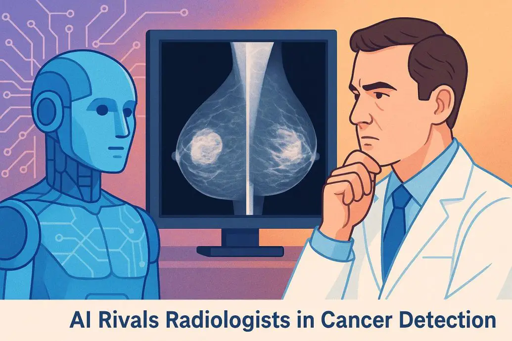
AI Rivals Radiologists in Cancer Detection
AI Rivals Radiologists in Cancer Detection highlights a pivotal advancement in diagnostic healthcare. Artificial intelligence has reached expert radiologist performance in detecting breast cancer using mammograms. Based on a comprehensive study published in Nature, this achievement was made possible by training a deep learning model on more than 1.2 million mammograms sourced from the U.S., U.K., and other international locations. The AI system effectively reduced false positives and unnecessary patient recalls, which are recurring issues in mammographic screening. While the results are promising, adoption in clinical practice will depend on rigorous validation, integration, and official regulatory approval. This development reinforces AI’s role as a clinical decision support system rather than a replacement for medical professionals.
Key Takeaways
- A deep learning algorithm trained on over 1.2 million mammograms matched expert radiologists in breast cancer detection performance.
- The AI significantly decreased false positives and unnecessary recalls, leading to more efficient diagnostics.
- Clinical implementation will require regulatory approval and seamless workflow integration.
- The model emphasizes AI’s capacity to support rather than replace radiologists.
Table of contents
- AI Rivals Radiologists in Cancer Detection
- Key Takeaways
- Global Context: The Urgency of Improved Breast Cancer Screening
- The Nature Study: Design, Dataset, and Model Architecture
- Performance Comparison: AI vs Radiologists
- How the AI Works: A Quick Glossary
- Radiologist Perspectives and Clinical Integration
- The Clinical Path Ahead: Validation and Regulation
- Comparing AI Models in Diagnostic Imaging
- Final Thoughts: Augmentation, Not Automation
- References
Global Context: The Urgency of Improved Breast Cancer Screening
Breast cancer remains the most diagnosed cancer and one of the leading causes of cancer-related deaths among women. According to the World Health Organization, over 2.3 million women were diagnosed with breast cancer in 2020, leading to approximately 685,000 deaths globally. Early and accurate screening drastically improves survival rates. Although mammography continues to be the standard tool for early detection, diagnostic inaccuracies persist. Studies show that false positives affect up to 10 percent of screenings and false negatives can account for 10 to 30 percent of missed cancers, which underscores a need for better diagnostic tools.
AI in medical imaging can improve diagnostic accuracy. Integrating AI in screening processes offers a promising path toward reducing errors and enhancing early detection across global healthcare systems. More insights can be found in this detailed look at AI in medical imaging diagnostics.
The Nature Study: Design, Dataset, and Model Architecture
The study from Nature represents an international collaboration involving DeepMind (now Google Health) and institutions from both the U.S. and U.K. The core of the research focused on developing a deep convolutional neural network that was trained on a curated dataset of over 1.2 million mammograms. This dataset included diverse patient demographics such as age, ethnicity, and breast tissue density to reflect real-world healthcare populations.
The model used supervised learning approaches. Annotated images confirmed by pathology were used to teach the system to recognize healthy versus malignant patterns within breast tissue. A separate and independent test set was used to benchmark the AI model against expert radiologists.
Performance Comparison: AI vs Radiologists
According to the study, the AI system reduced false positives by 5.7 percent and false negatives by 9.4 percent in the U.S. dataset. In the U.K. dataset, the false positive rate dropped by 1.2 percent and the false negative rate by 2.7 percent. These results demonstrate the AI’s consistent ability to improve diagnostic performance for breast cancer detection across varied populations.
Unlike human diagnosticians, who may differ in interpretation, the AI provided consistent evaluations. Variability among radiologists had already been well documented, especially in ambiguous cases. The study illustrates how AI can bring standardization and reliability to the reading process.
Table: Diagnostic Accuracy Comparison
| Model/Reader | False Positive Rate | False Negative Rate | Population Tested |
|---|---|---|---|
| DeepMind AI Model | -5.7% (US) / -1.2% (UK) | -9.4% (US) / -2.7% (UK) | 1.2M+ Mammograms (US, UK) |
| Radiologists (Avg.) | Standard Baseline | Standard Baseline | Identical control group |
| CheXNet (Chest Imaging AI) | Not directly comparable | Improved pneumonia diagnosis | Chest X-ray dataset |
How the AI Works: A Quick Glossary
Here is a simplified glossary for readers who are not familiar with the technological terms involved
- Deep Learning: A type of machine learning that uses neural networks to process complex data patterns, particularly in images.
- Convolutional Neural Networks (CNNs): A type of neural network specially suited for image classification and pattern recognition.
- Mammogram: An imaging method that uses low-dose X-rays to examine breast tissue.
- False Positive: A test result that indicates cancer when none is present.
- False Negative: A test result that fails to detect existing cancer.
Radiologist Perspectives and Clinical Integration
Medical professionals largely view AI as a valuable adjunct. Dr. Constance Lehman, chief of Breast Imaging at Massachusetts General Hospital, noted in a CNN interview that reproducible decision-support tools are needed, not replacements for expert judgment.
AI could assist in double reading scenarios, offering a second opinion or flagging complicated cases. Successful integration will depend on compatibility with existing systems like PACS and EMRs to ensure workflow efficiency. These are the practical steps required for AI and radiologists to collaborate effectively toward better patient outcomes.
The Clinical Path Ahead: Validation and Regulation
Despite the impressive findings, real-world implementation entails several steps. Regulatory agencies such as the U.S. FDA and the U.K. MHRA must evaluate the system for safety, reliability, and effectiveness. Multiple pilot programs are underway to test AI performance in live hospital settings and gather feedback on usability and reliability.
These trials aim to address concerns related to liability, patient safety, algorithmic bias, and data privacy. Transparency in the AI’s decision-making process is another critical factor for building clinical trust. This process mirrors developments already seen in AI-powered cancer treatment advancements.
Comparing AI Models in Diagnostic Imaging
Diagnostic AI models have already demonstrated success across various medical domains. Some examples include:
- CheXNet: Created by Stanford for diagnosing pneumonia from chest X-rays.
- PathAI: Uses computer vision to assess digital biopsy slides for cancer pathology.
- DeepMind Eye Disease Model: Developed to detect more than 50 retinal disorders with hospital-level accuracy.
These examples illustrate how AI is becoming a cross-disciplinary aid within modern healthcare. To explore more applications, see the overview of AI-driven innovations in healthcare.
Final Thoughts: Augmentation, Not Automation
AI’s growing role in breast cancer detection marks a significant shift in healthcare. It aims to enhance, not replace, human expertise by offering greater consistency and reducing diagnostic errors. Ethical deployment, strict regulations, and thoughtful integration will dictate how effectively AI complements clinical operations. As AI continues to mature, it has potential to reshape diagnostic medicine in a manner that prioritizes patient outcomes while preserving professional oversight. To understand more about the wider implications, visit this feature on the impact of artificial intelligence in healthcare.
References
- McKinney, S. M., Sieniek, M., Godbole, V., Godwin, J., Antropova, N., Ashrafian, H., … & Suleyman, M. (2020). International evaluation of an AI system for breast cancer screening. Nature</em