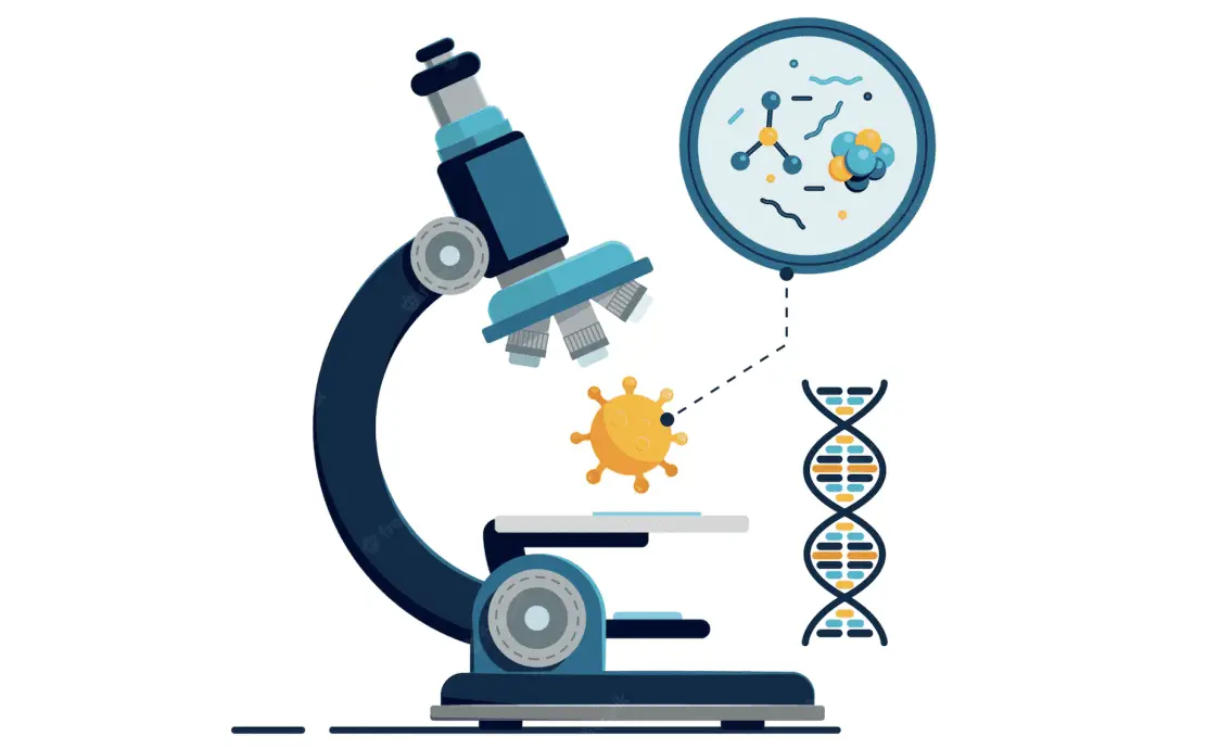
In recent years, automated microscopy has risen to prominence, particularly for managing complex and sophisticated applications. These applications often necessitate continuous and repetitive observations, which can be an arduous and time-consuming task for human operators. The time commitment alone can span several hours, making it a challenging endeavor for individuals.
Advancements in automated microscopy technology have led to marked improvements in performance, outpacing its traditional counterparts. The system now efficiently tackles labor-intensive activities and processes that used to consume significant amounts of time. From focus control to image acquisition, automation now handles a multitude of tasks that previously required manual intervention. This article aims to provide a comprehensive understanding of automated microscopy. It delves into its numerous aspects, offering valuable insights into its capabilities, advantages, and the ways it is revolutionizing scientific research and data collection.
Table of contents
Also Read: Is robotics computer science or engineering?
What Is The Definition Of Automated Microscopy
Automated microscopy refers to the use of computer-controlled systems to perform microscopy functions without the need for manual operation. It integrates software, hardware, and advanced algorithms to streamline tasks like image capturing, focusing, and data analysis. This allows for high-throughput data collection, real-time analysis, and often yields more consistent and reliable results.
What Is Automated Microscopy?
Technology has advanced, and so has microscopy. This progress lets you easily integrate high-tech features from other sectors to streamline data collection. Changes in microscope components now support automation, yielding compelling results.
Automation in microscopy offers several advantages in data acquisition, including instant data archiving from real-time analysis. It also maintains samples in sterile conditions. These conditions can be imbalances or extreme temperatures like those in carbon dioxide. The microscope captures and visualizes samples without human intervention. The hardware stays consistent, while the researcher collects data from a safe distance.
The concept of automated microscopy began in the 1960s at Cambridge. Researchers developed an analyzer for detecting microscope images. The technology took time to mature and compete with traditional methods. Yet it did evolve, bringing significant changes to scientific research. Digital elements in traditional microscopes were game-changers.
Using cutting-edge automation and artificial intelligence in microscopy helps in efficient data collection and system maintenance. This reduces both time and effort to achieve reliable results.
What Is Traditional Microscopy?
In labs, you often see traditional microscopes. These require manual operation. Users handle tasks like focusing, slide placement, and image capture themselves. They also manually adjust settings such as lighting and magnification. Data collection and analysis depend on human input. This manual process can be time-consuming and prone to errors. Despite these limitations, traditional microscopes remain valuable for specific applications. They provide a hands-on approach that some researchers prefer.
Microscopy In The Current Day
When you are looking at a modern-day microscope, you can have different types of optical components that you can optimize. These components include focus control, light sources, stages, filter wheels, shutters, and many more.
These components are replaced by electronic ones and controlled by intuitive imaging software. Precisely, the automation of how microscopes can capture images automates the whole workflow. As such, the process requires human intervention after the images are produced.
Automated microscopy is useful when it comes to using the same in applications that need a higher number of continuous observations spread over a definite period. Therefore, whether it is about highly throughput analysis or live cell imaging, science professionals use automated microscopy to uncover more information than ever before.
Whether you use them for point-of-care or research-based, modern-day microscopes have components that are automated. Even if you want to transform your traditional microscopy into an automated one, you can do that effectively simply by changing the components. Next, connect them to a computer and control them with the available software package.
Still, if you wish to transform an automated imaging system that works optimally, it might be a complex task. Accomplishing the task involve experience in electronics and optics, many years of experience, and plenty of time. As such, it makes sense why purchasing a fully-functional automated system is the most effective way in this world full of possibilities.
So, whether you buy a readymade one or build one, a proper understanding of the components of automated microscopy helps you to decide which system might be better for the research work.
How Does Automated Microscopy Work?
Automated microscopy offers significant advantages over traditional methods, particularly in focus and stage control. By utilizing focus motors on transmission gears, you can achieve precise control over the microscope’s focal point. This exactitude is imperative for capturing sharp, detailed images. To enhance this capability, many systems integrate advanced image acquisition software designed specifically for autofocusing tasks. The software often incorporates sophisticated algorithms that continuously adjust focus parameters in real time. This feature not only improves the accuracy of the data but also minimizes the risk of human error, making the microscope highly efficient in handling automated, intricate imaging tasks. As a result, researchers can allocate their attention to data interpretation and other critical aspects of their work.
When it comes to selecting wavelengths, traditional methods often involve the use of acousto-optic tunable filters, monochromators, beam splitting units, and filters. These methods are not only slow but also require constant human intervention, making them less efficient. As an alternative, motor-rotated filter wheels enable rapid and accurate wavelength selection, thus streamlining the process and freeing up operator time.
The flexibility of automated settings is another noteworthy feature. These settings allow for experiments to run without constant oversight. Automated microscopy can control various other aspects such as image acquisition, environmental conditions, and illumination sources. This multi-faceted automation expands the effectiveness and applications of microscopy in scientific research.
In contrast to traditional microscopy, which demands extensive manual operation, automated systems require minimal human input. This technological leap fundamentally changes the landscape of scientific research, creating new possibilities and opportunities for more complex and nuanced studies.
Also Read: Can AI and Machine Learning Simulate the Human Brain?
Benefits Of Automated Microscopy
Automated microscopy improves the process of data collection and handles the sample in easy steps so that it remains free of inconsistencies in the final measurement. Automation provides an ultimate solution to several issues so that the sample analysis is conducted with minimal human intervention. Here are the benefits of the system so that you can know everything about it.
Automation Can Increase Productivity
There is no denying that analysts perform a variety of tasks in the laboratory other than sample preparation and data collection. The other tasks might include data analysis, experiment design, collaborating with other researchers, and so on.
Automated microscopy streamlines high-level tasks that are usually tedious and time-consuming. It not only facilitates unattended operation but also incorporates pre-set analysis protocols. These protocols automatically handle preprocessing and evaluation of samples. While the automated system takes care of these functions, users are free to perform other crucial tasks. This multitasking capability significantly enhances lab efficiency and data reliability.
Automation Improves Repeatability
Analytical measurements involve sampling handling tasks such as cooling, heating, mixing, and injecting the samples inside the device. Because these steps are done manually, it gives you inconsistent results.
The worst part is that the variabilities could result in underlying differences between those samples. Now, sample handling steps such as mixing and dilution could give rise to factors like particle content if they aren’t performed carefully.
Automation can take good care during the sample handling period. As such, it results in high-quality information from the instrument that is valuable in many research applications and contexts.
Integrated Software Makes The Task Easy
Automated microscopy offers a user-friendly experience if both the software and hardware are easy to operate. Researchers developing analytical protocols benefit from a well-designed, robust interface. This interface provides straightforward access to all necessary functions. Simplicity in operation is particularly beneficial during routine analyses. In these instances, researchers often prefer an interface that is streamlined and intuitive.
With automated microscopy, executing complex analyses becomes as easy as clicking a button. This ease of use can lead to greater efficiency in the lab, enabling researchers to complete more analyses in less time. Moreover, a simplified but powerful user interface minimizes the likelihood of errors, enhancing data reliability. Overall, the user-centric design of automated microscopy systems improves not just operational efficiency, but also the quality of scientific research. This contributes to accelerating discoveries and innovations in various scientific fields.
Software and Hardware Adaptability For User’s Need
For a successful integration into an existing workflow, it would depend on the flexibility of the automation in terms of the software and hardware. However, certain factors like options and size of the sample deck layout determine the exact number of samples and the processing steps that the instrument can automate.
Similarly, the software used on the instrument should allow the users to come up with automated protocols so that they can utilize the different types of functionality that the liquid handler includes.
Remote Working
Automated microscopy incorporates advanced technology that allows for cloud-based connectivity. By linking the microscope to a cloud system, it becomes accessible for remote operation. Researchers can control the device, adjust settings, and analyze samples through a computer interface. This can happen even if the researcher is miles away from the actual lab equipment. This remote capability is not just convenient; it also revolutionizes the way research is conducted.
It breaks down geographical barriers and enables collaborative efforts across different locations. Researchers no longer need to be physically present in the lab to conduct experiments or collect data. This saves time and resources, and increases the flexibility and efficiency of scientific investigations. Additionally, cloud storage facilitates seamless data sharing and archival, making it easier to manage large datasets. In summary, the cloud compatibility of automated microscopy enhances research scope, operational flexibility, and data management, all while allowing researchers to focus on other important tasks.
Brings Maximum Productivity
No wonder, a complete solution can bring maximum productivity to any setup, and biotherapeutics are not an exception. In addition, long-term association and success with different types of analytical measurements depend on an automated instrument.
However, it depends on the proper system maintenance and prompt service as well. Therefore, the provider of an automated and integrated solution must provide after-sales service.
There should be comprehensive support for the whole setup without compromising on the overall functionalities and quality. As a result, these services help to maximize uptime and improve productivity to a great extent.
Conclusion
Automated microscopy involves working with a device that interacts with several components and intuitive software solutions. The device is used for staging photos in a different magnification and lighting conditions. While you are looking for solutions to meet your requirement, make sure that you choose components that are compatible with each other and deliver sheer performance to make sure that you didn’t disrupt the process.
